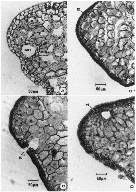
Fig. 1 from Moss et al. (1998): “(A–D) Light micrographs of transverse sections of first-year red spruce needles, collected at the mid-elevation site on Mt. Moosilauke, showing normal developmental maturation over the 1988 growing season. E, epidermis; H, hypodermis; RC, resin canal; S, stomate. (A) Collected June 29, 1988. Note immature light staining of epidermal tissue system and numerous discrete chloroplasts in mesophyll. (B) Collected August 2, 1988. Note dark staining, thick-walled epidermal system and still immature mesophyll. (C) Collected August 17, 1988. Note fully developed mesophyll with discrete chloroplasts characterizing a healthy needle. (D) Collected September 20, 1988. Note discrete chloroplasts and fine granular, light staining of cytoplasmic contents. Also note absence of damage symptoms.”
 The Botanist in the Kitchen